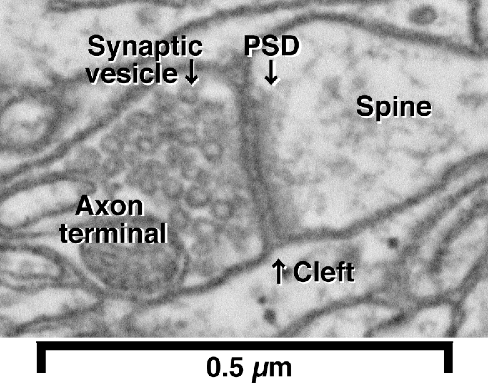
A high magnification image of synapse obtained by electron microscopy
Por um escritor misterioso
Last updated 28 março 2025

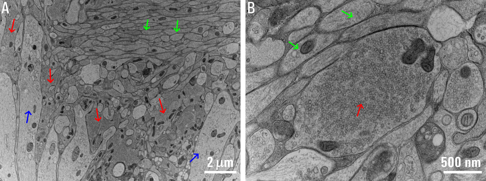
Investigating Synapses in Brain Slices with Enhanced Functional

Targeting Functionally Characterized Synaptic Architecture Using

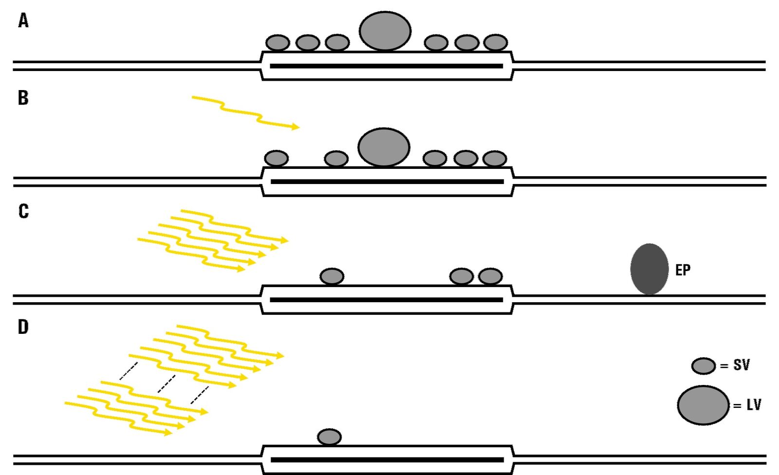
Discovery of a New Mechanism Regulating Information in the Brain
NIPS_Research
Investigating Synapses in Brain Slices with Enhanced Functional
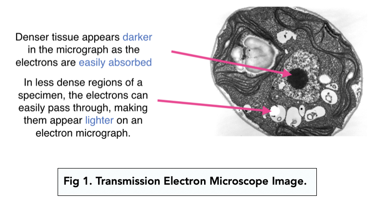
Studying Cells: Electron Microscopes (A-level Biology) - Study Mind

Choosing the Right Scanning Electron Microscope for Your

Under a high magnification of 10, 000x, this scanning electron
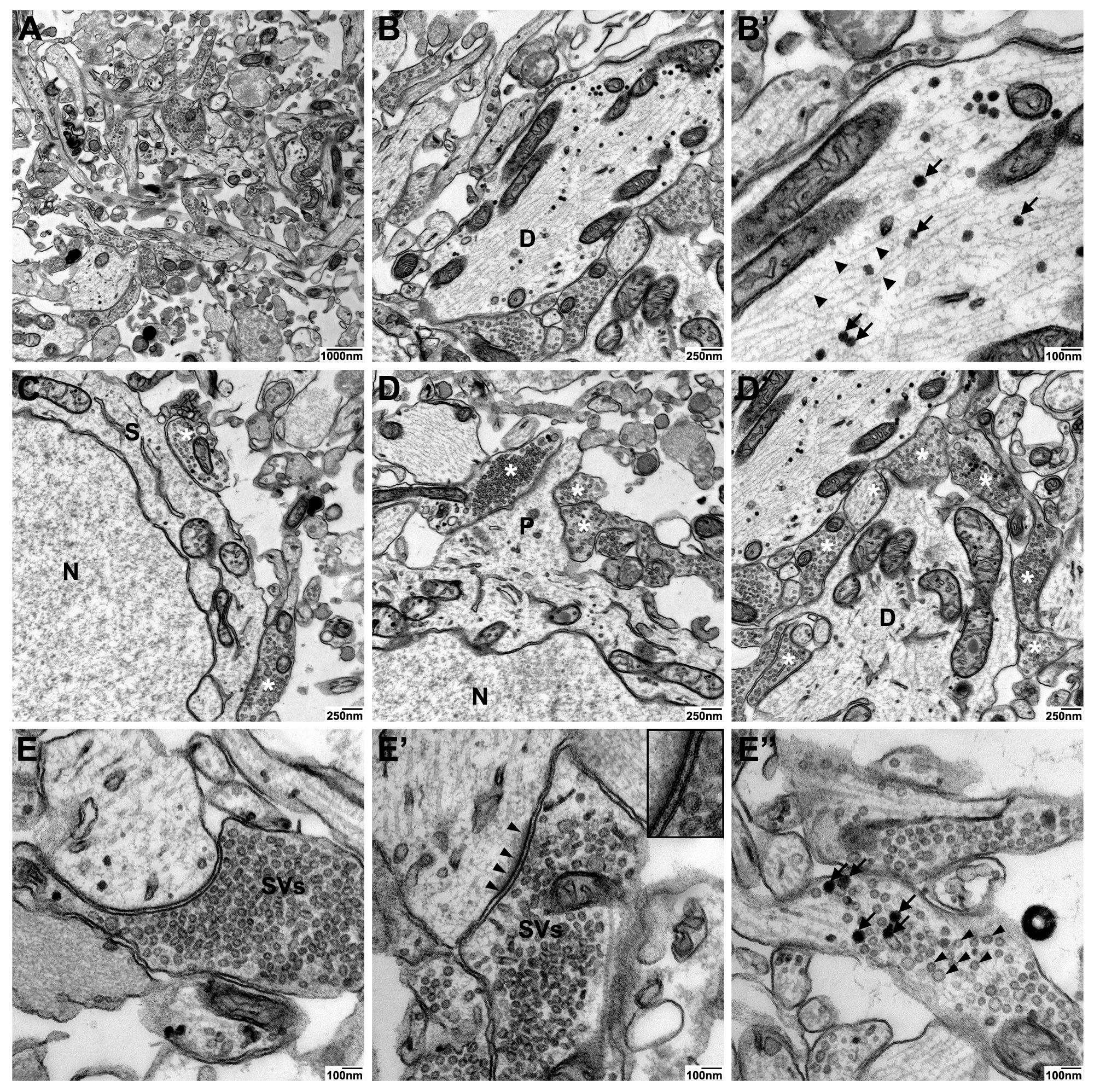
IJMS, Free Full-Text
High Magnification Electron Micrograph of a Typical Synapse In the
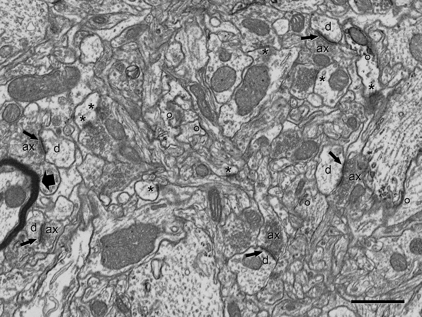
Frontiers Counting synapses using FIB/SEM microscopy: a true

Automated Detection and Localization of Synaptic Vesicles in
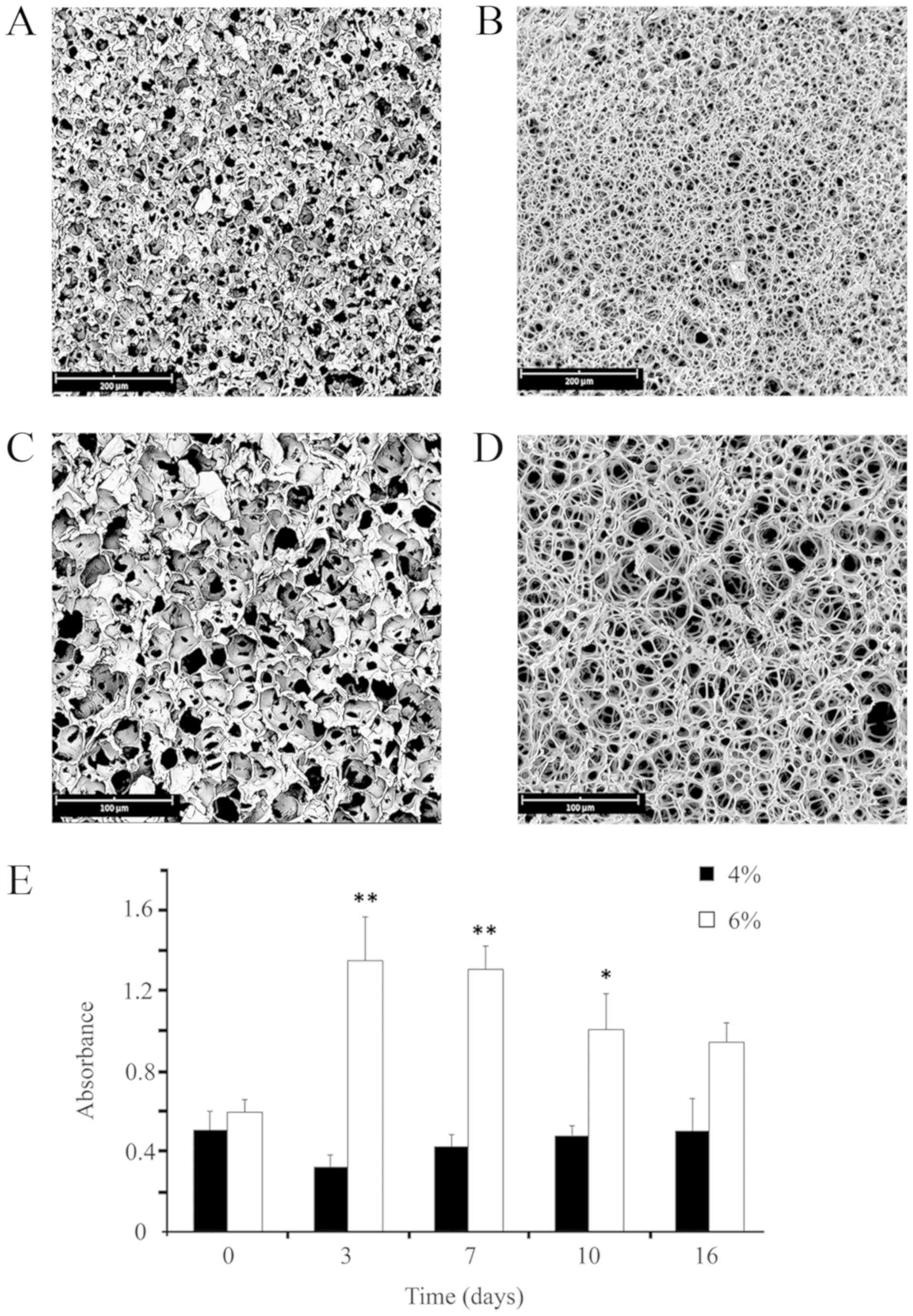
A 3D‑scaffold of PLLA induces the morphological differentiation
Recomendado para você
-
 R.I.P Synapse X (2016 - 2023) : r/ROBLOXExploiting28 março 2025
 -
 Synapse X Server Status Check Synapse X Is Currently Down for Maintenance28 março 2025
 -
X Public Data (Twitter) Connector - Supermetrics28 março 2025
-
 Peer support for people with a brain injury28 março 2025
 -
 This Week in Matrix 2021-12-1728 março 2025
 -
 BitAntiCheat - A server-sided, general purpose anti-cheat! - Community Resources - Developer Forum28 março 2025
 -
 There is a way to set up Synapse Administrators with a BICEP module? - Stack Overflow28 março 2025
 -
 Engineered adhesion molecules drive synapse organization28 março 2025
 -
 Why Is Synapse X Is Not Currently Not Available For Purchase & What to Do Now?28 março 2025
 -
 Razer Synapse: What it does, and how to use it28 março 2025
você pode gostar
-
 Boneca Bebê Reborn Realista Menina De Silicone 42cm Cheirosa28 março 2025
 -
 Dragon Ball Super Chapter 88 Delayed As Manga Goes On Hiatus28 março 2025
 -
 Linkle Mod: Wind Waker HD [The Legend of Zelda: The Wind Waker HD28 março 2025
 -
 15+ EASY Hors d'oeuvre Ideas Your Party Needs! - Aleka's Get-Together28 março 2025
 -
 Cadeira de Barbearia Princess Marri - Ponto do Cabeleireiro28 março 2025
 -
 File:Antigua tribuna del estadio de Ferro Carril Oeste.jpg28 março 2025
 -
 Caminhão Hot Wheels Might K Hcw70 Rosa C28 março 2025
 -
 Pin de Gee Pin em Muñequitas Enfeites de biscuit, Biscuit, Bonecos de biscuit28 março 2025
 -
![Livro Diary Of Mike The Roblox Noob: Murder Mystery 2, Jailbreak, Meepcity - Mike, Roblox [2017]](https://http2.mlstatic.com/D_NQ_NP_790788-MLB69568591707_052023-O.webp) Livro Diary Of Mike The Roblox Noob: Murder Mystery 2, Jailbreak, Meepcity - Mike, Roblox [2017]28 março 2025
Livro Diary Of Mike The Roblox Noob: Murder Mystery 2, Jailbreak, Meepcity - Mike, Roblox [2017]28 março 2025 -
 Bazzi - Paradise (Lyrics)28 março 2025
