CT brain image gallery - SAH
Por um escritor misterioso
Last updated 31 março 2025
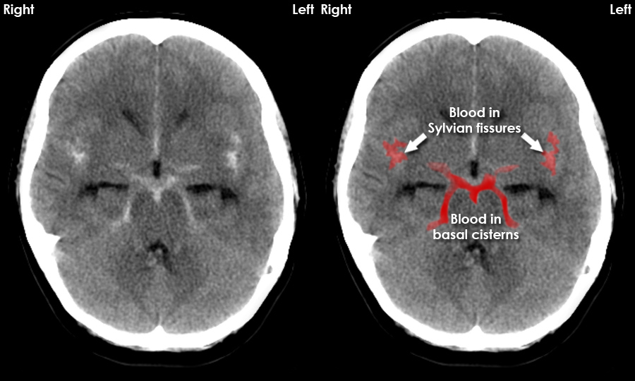
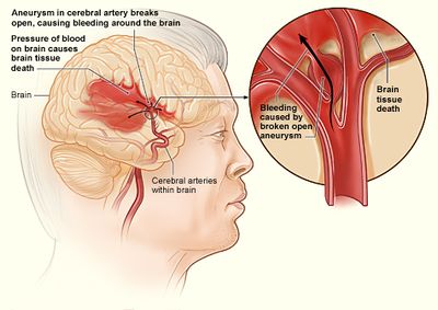
CT brain images - example of subarachnoid haemorrhage as seen on CT. Dense material in the basal cisterns, fissures and sulci represents acute bleeding into the subarachnoid space.

Figure 3 from Hemorrhage slices detection in brain CT images

Figure 69-5 from How to Read a Head CT Scan

How to identify common causes of subarachnoid hemorrhages on comp

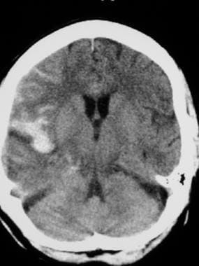
Brain computed tomography (CT); Diffuse subarachnoid hemorrhage

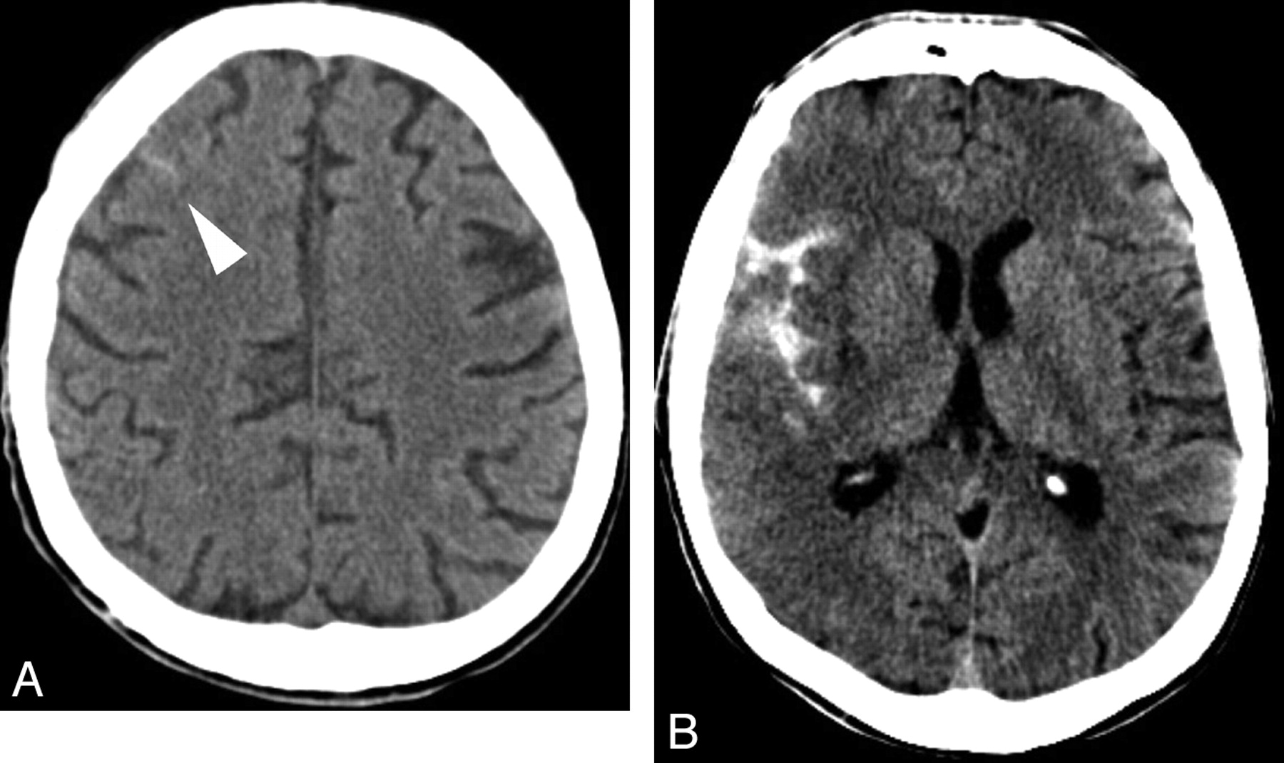
Axial brain CT scans obtained on admission, showing an SAH with

Subarachnoid Hemorrhage (Chapter 13) - Textbook of Stroke Medicine
Subarachnoid Hemorrhage Workup: Approach Considerations, Computed
Isolated Acute Nontraumatic Cortical Subarachnoid Hemorrhage

Subarachnoid Hematoma (SAH) - Trial Exhibits Inc.

120+ Subarachnoid Hemorrhage Stock Photos, Pictures & Royalty-Free
Brain MRI: How to read MRI brain scan

How to identify common causes of subarachnoid hemorrhages on comp
Recomendado para você
-
 Sah, MSM The Lost Landscapes Wiki31 março 2025
 -
FIGURE. Diagnosis and treatment in 284 consecutive patients with31 março 2025
-
 Jay Sah, College of Arts, Sciences & Education31 março 2025
 -
 Subarachnoid Hemorrhage (SAH) - Physiopedia31 março 2025
 -
 Subarachnoid Hemorrhage (SAH) - Willis-Knighton Health System31 março 2025
 -
 SAAF™ Side Access Housings (SAH) - AAF International31 março 2025
 -
SAH Trauma-Informed Embodied Spirituality (@sahdsimone31 março 2025
-
 Sah Sultan, Magnificent Baddie Wiki31 março 2025
 -
 Posso aceitar amostra grátis de Sáh Assumpção - Pensador31 março 2025
 -
 UFMG - Universidade Federal de Minas Gerais - Eventos31 março 2025

você pode gostar
-
TUTORIAL COMO PEGAR TODOS OS TROFÉUS - DOODLE ILHA DOS CAMPEÕES (PC) AGORA NO IGTV. Tutorial / Guia para ajudar a gatinha Lucky a pegar / obter / conseguir todos os31 março 2025
-
 Unable to upload any new clothings? - Art Design Support - Developer Forum31 março 2025
 -
roblox-cookie-logger · GitHub Topics · GitHub31 março 2025
-
 Haruna (Kore Wa Zombie Desu Ka?) (Cosplay)31 março 2025
 -
:quality(75)/cloudfront-us-east-1.images.arcpublishing.com/elcomercio/ILUFBJRWIBDT5A2J5BRRCORYFQ.jpg) Sigue ahora en vivo online, Chile 1-1 Uruguay: sigue ahora el partido de hoy por las Eliminatorias 2022, Partido de hoy, fútbol en vivo, INTERNACIONAL31 março 2025
Sigue ahora en vivo online, Chile 1-1 Uruguay: sigue ahora el partido de hoy por las Eliminatorias 2022, Partido de hoy, fútbol en vivo, INTERNACIONAL31 março 2025 -
 Gohan Beast Anime dragon ball goku, Anime dragon ball super, Anime dragon ball31 março 2025
 -
 Um Grupo De Pessoas Jogando Futebol De Vetor PNG , Jogando Futebol Clipart, Luzes, Atleta Imagem PNG e PSD Para Download Gratuito31 março 2025
 -
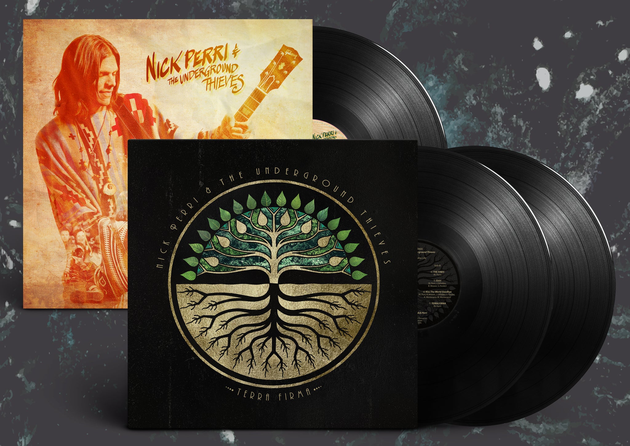 Full Catalog – Nick Perri Music31 março 2025
 -
 Wormate.io 🕹️ Jogue no CrazyGames31 março 2025
 -
Máquina Pop31 março 2025

