Figure 1 from Brain surface temperature under a craniotomy.
Por um escritor misterioso
Last updated 28 março 2025

Fig. 1. Rapid cooling of the brain surface in an in vivo mouse preparation. A: schematic representation of a cranial window during recording of temperature and single-cell activity in the anesthetized mouse. The main potential routes of heat transfer are indicated. B: brain surface temperature measured with the thermocouple during replacement of the artificial cerebrospinal fluid (ACSF) with fresh ACSF warmed to 38°C. ACSF was replaced twice, indicated by the arrowheads. - "Brain surface temperature under a craniotomy."

Cranial imaging window implantation technique for longitudinal multimodal imaging of the brain environment in live mice - ScienceDirect
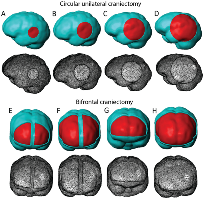
Decompressive craniectomy of post-traumatic brain injury: an in silico modelling approach for intracranial hypertension management
Brain Sciences, Free Full-Text

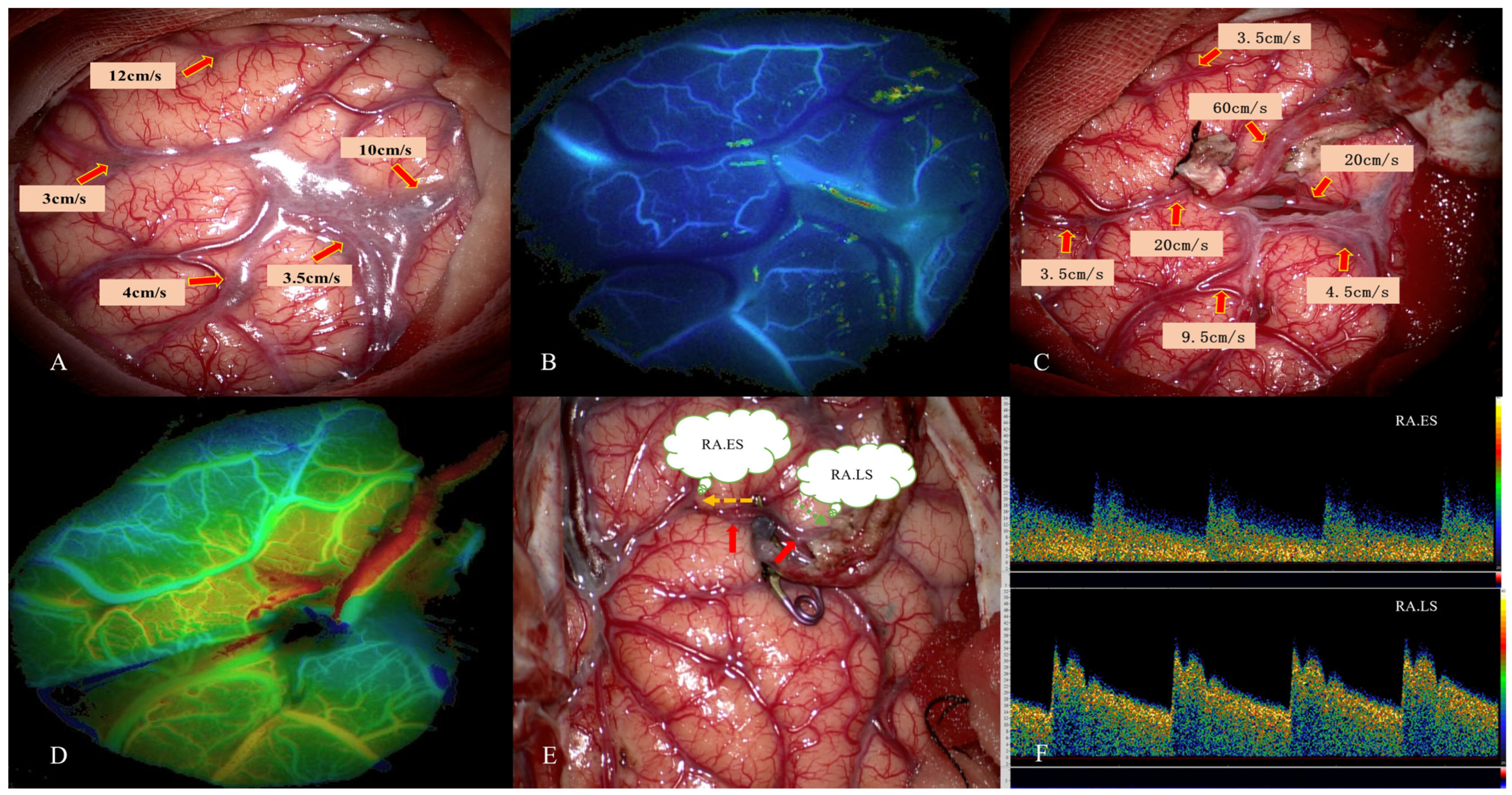
Craniotomy for acute monitoring of pial vessels in the rodent brain - ScienceDirect
JCM, Free Full-Text
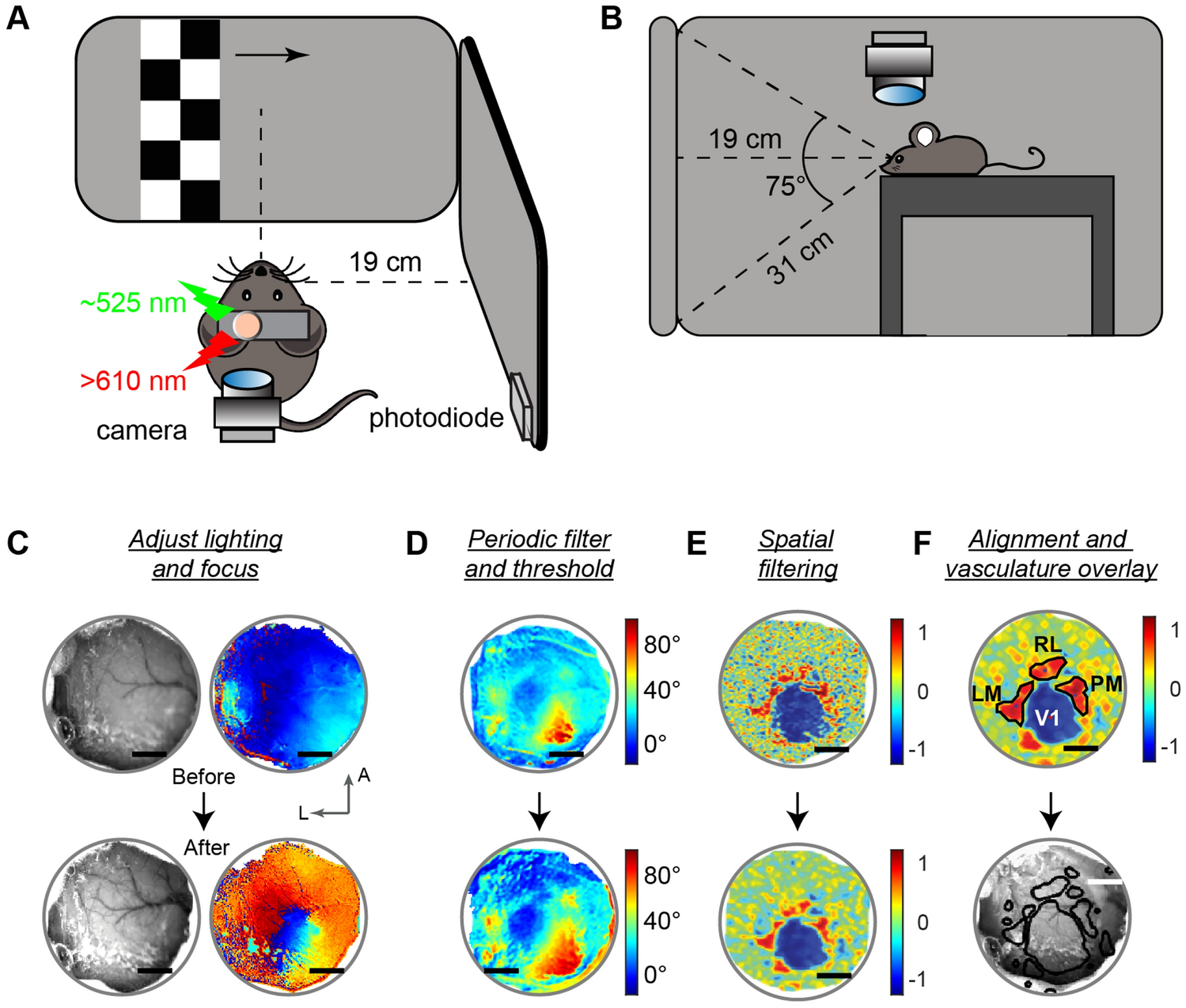
Optimizing intact skull intrinsic signal imaging for subsequent targeted electrophysiology across mouse visual cortex
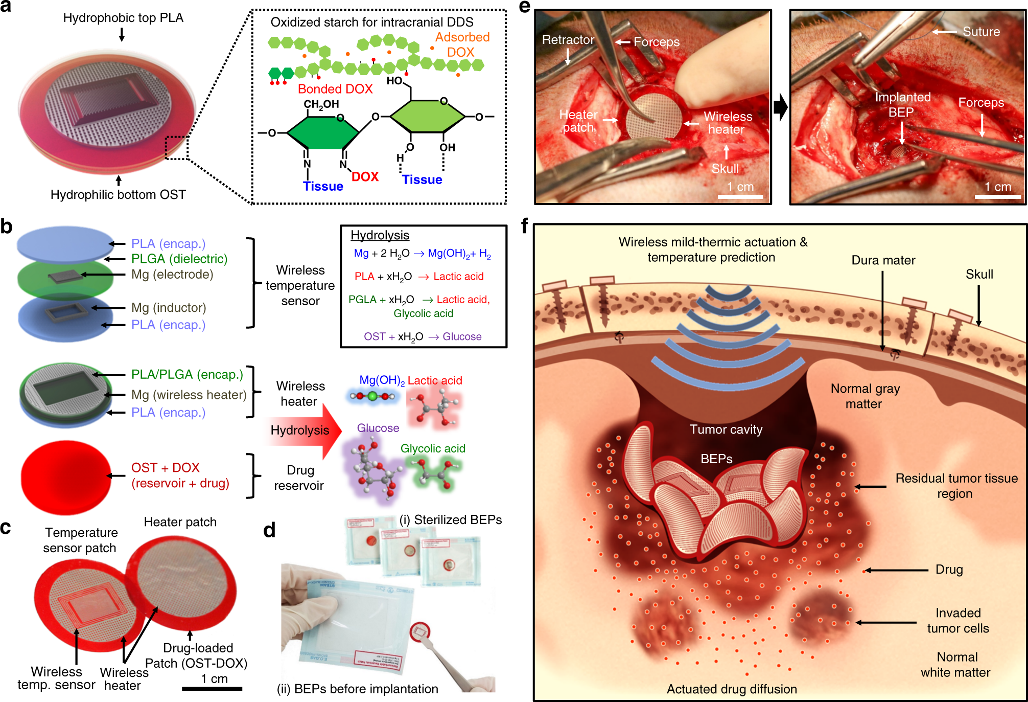
Flexible, sticky, and biodegradable wireless device for drug delivery to brain tumors

Figure 1 from Brain surface temperature under a craniotomy.

PDF] Jugular bulb temperature: comparison with brain surface and core temperatures in neurosurgical patients during mild hypothermia.
JCM, Free Full-Text

Brain surface temperature under a craniotomy

Photothrombotic Middle Cerebral Artery Occlusion in Mice: A Novel Model of Ischemic Stroke

Brain temperature but not core temperature increases during spreading depolarizations in patients with spontaneous intracerebral hemorrhage - Alois J Schiefecker, Mario Kofler, Max Gaasch, Ronny Beer, Iris Unterberger, Bettina Pfausler, Gregor Broessner

Longitudinal two-photon calcium imaging with ultra-large cranial window for head-fixed mice - ScienceDirect

Temporal/Subtemporal Craniotomy
Recomendado para você
-
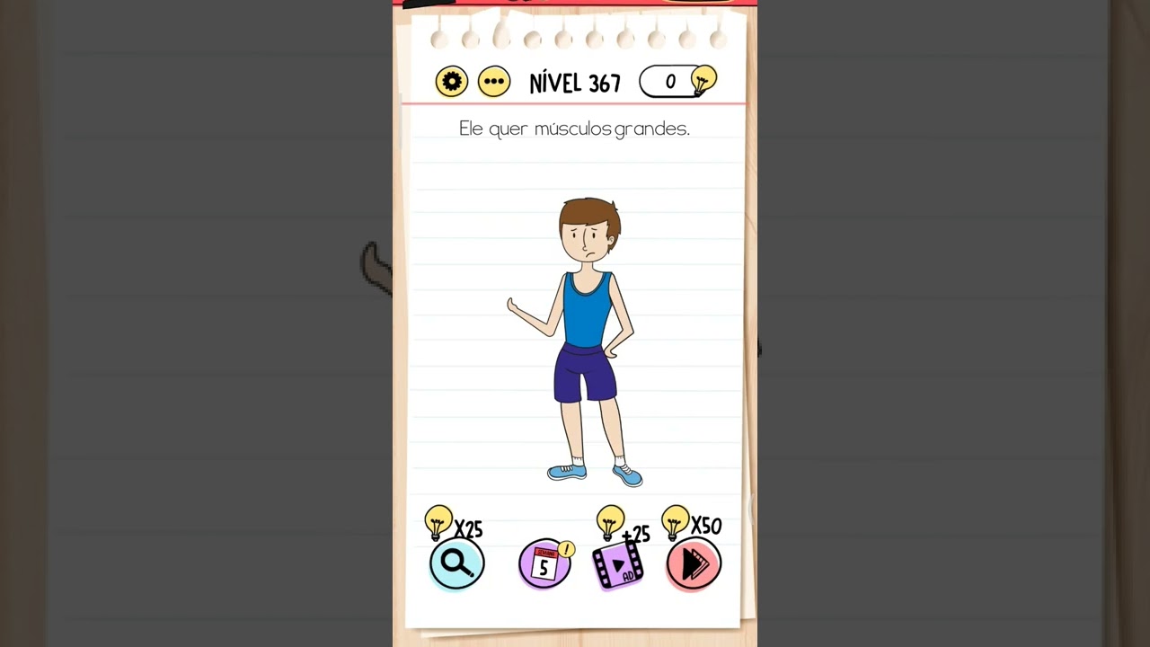 BRAIN TEST NÍVEL 367 EM PORTUGUÊS28 março 2025
 -
Death Incoming (level 367) #Android #Game #gameplay #gaming #apk28 março 2025
-
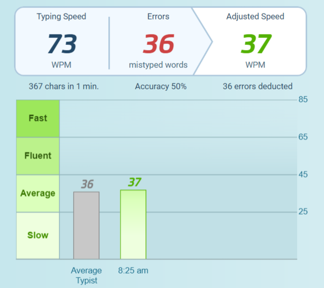 Tech Thursday: Voice-to-Text - by Breana Bayraktar28 março 2025
 -
d-Amino Acid Levels in Perfused Mouse Brain Tissue and Blood: A Comparative Study28 março 2025
-
 Association of body mass index and waist-to-hip ratio with brain structure28 março 2025
 -
 Use of MRI in the diagnosis of fetal brain abnormalities in utero (MERIDIAN): a multicentre, prospective cohort study - The Lancet28 março 2025
 -
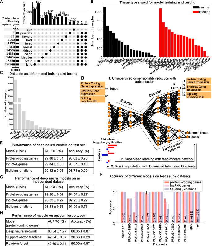 Identifying common transcriptome signatures of cancer by interpreting deep learning models, Genome Biology28 março 2025
 -
 Saatnya mencari cuan! (Brain Test Level 367) - CadeMedia28 março 2025
 -
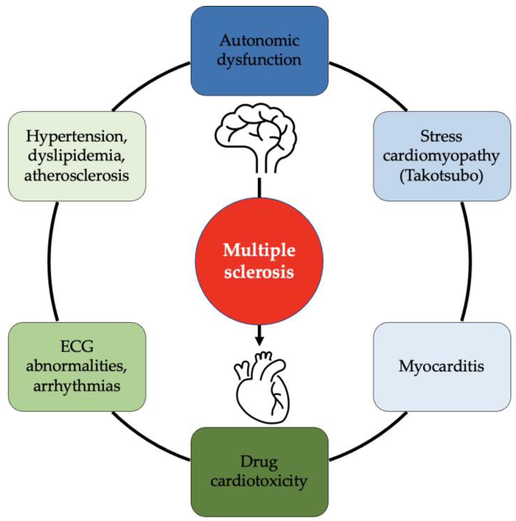 JCDD, Free Full-Text28 março 2025
 -
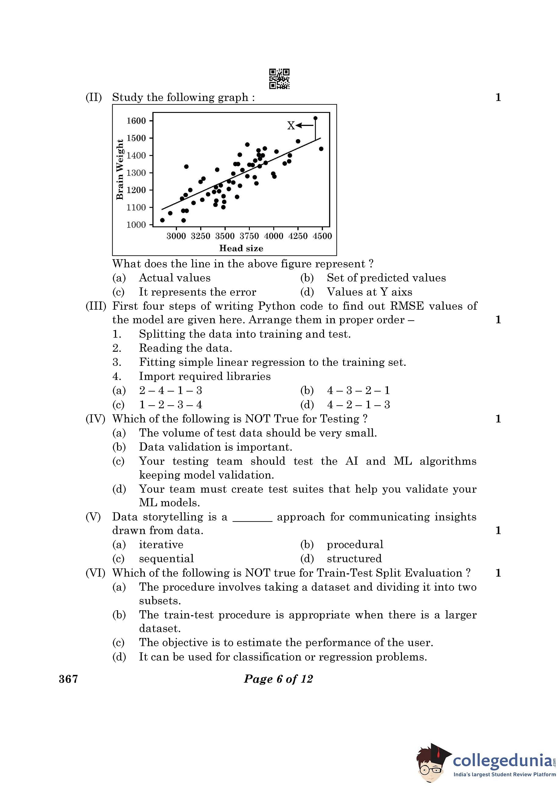
CBSE Class 12 Artificial Intelligence Question Paper 2023 with Answer Key (February 22, Set 4 - 367)28 março 2025


você pode gostar
-
 Is Resident Evil 3 remake coming to Nintendo Switch?28 março 2025
 -
25% OFF - P2design Academy - Black Friday - p2design28 março 2025
-
The Man from the window chap 2 APK (Android Game) - Free Download28 março 2025
-
 Sakamoto desu ga - 08 -18 - Lost in Anime28 março 2025
 -
 Class 1 - Parts of a Computer - Cyber Square28 março 2025
 -
 Turgut Bey ölüyor mu? Kuruluş Osman'ın Turgut Alp'i Rüzgar Aksoy diziden ayrılıyor mu? - Aydın Haber, Son Dakika Aydın Haberleri28 março 2025
 -
 Share Memes, Make Memes, Make Money, Make Communities28 março 2025
 -
 project slayers helping server|TikTok Search28 março 2025
 -
Super Mario™ 3D World + Bowser's Fury for Nintendo Switch - Nintendo Official Site28 março 2025
-
 Battlefield 2042 atinge o maior número de jogadores desde o lançamento28 março 2025